Ear Disorders
CLICK ON A TOPIC BELOW TO LEARN MORE
Deafness
Deafness can be partial or complete, temporary or permanent. A complete, permanent lack of hearing may be congenital (present at birth) in white animals and certain breeds (eg. Dalmations). Other causes of deafness include degeneration with aged pets, severe nutritional disorders, immune disorders, chronic ear infections, trauma and toxins (eg. aminoglycosides). Unfortunately there may be no cure for many types of deafness, and the hearing loss is generally permanent. Fortunately, total deafness is rare. A partial hearing loss may also be permanent. Laboratory assessment of hearing is afforded through the BAER (brainstem auditory evoked response) test. Deafness, the result of infection, may respond to treatment.
Ear Mite Infestation
Ear mites are microscopic parasites that live in the ear canals of dogs and cats. These mites are highly contagious and frequently infest entire litters of puppies and kittens. Signs include dark discharge (“coffee ground-like”) within the ear, scratching at the ears, redness or blood within the ear and head shaking. Diagnosis is afforded through microscopic exam of the ear discharge. Treatment includes ear cleaning, ear flushes, miticidal drops, topical treatment (eg. selamectin) and parental ivermectin. If more than one dog or cat is present in the home, and one is found to be infected, then all should be carefully examined for ear mites and treated either as above or prophylactically.
Hematoma of the Ear Flap (aural hematoma)
Aural hematoma is an accumulation of blood between the cartilage and skin of the ear flap. The lesion is a result of a broken blood vessel caused by vigorous head shaking, scratching at the ears with the back feet or other trauma. Underlying causes include ear infections, ear mites and fleas. Signs of the lesion include swelling of the ear flap which may be warm and sore. Diagnosis is afforded via physical exam, aspiration and cytology. Treatment includes drainage, pressure bandages and/or surgery. Underlying causes should also be treated.
Otitis Externa
Otitis externa is an inflammation of the external ear canal. Causes include impaired drainage of the ear, neoplasia, bacteria, fungi, ear mites, accumulation of wax, thick or matted hair in the ear canal, debris, and trauma. The ears of dogs (especially those with pendulous ears) and cats are ideal for the growth of bacteria and fungi because they are moist and warm, and may contain wax and other debris. The dead end, funnel shape configuration of the ear canal effectively traps debris and further complicates treatment of infections. Signs of otitis externa include discharge within the ear, scratching at the ears, redness or blood within the ear, malodor and head shaking. Diagnosis is afforded through accurate history, physical exam and microscopic exam of the ear discharge as well as complete blood count, blood chemistries, cytology, microbe sensitivity tests, skin scrapes, fungal tests and intradermal skin testing . Treatment may include ear cleaning, ear flushes, topical treatment, parental (eg. via injection) treatment, immune therapy (eg. hyposensitization), anti-inflammatories, antibiotics, antifungals, antiparasitics and surgery (eg. lateral ear resection).
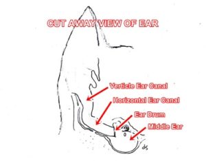
Otitis Media
Otitis media is inflammation/infection of the middle ear. It usually results from infection of the external ear canal or pharynx (back of mouth) spreading into the middle ear. Causes include bacteria, fungi, viruses, periodontal disease, neoplasia, foreign bodies, debris, ulceration or trauma (eg. improper ear cleaning that has ruptured the ear drum, allowing bacteria to reach the middle ear). Signs of middle ear infections include odor, discharge, ear scratching, pain, head shaking and vomiting, anorexia, facial nerve injury (drooping of the upper lip, drooling, decreased blinking on the side effected) and head tilt. Diagnosis is afforded by history, physical exam, complete blood count, blood chemistries, otoscopic exam, skull x-rays, cytology, culture and sensitivity. Treatment may include anti-inflammatories, antibiotics, antifungals, dental cleaning, ear flushing and surgery.


