Muscular Skeletal Disorders
CLICK ON A TOPIC BELOW TO LEARN MORE
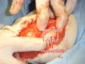
Arthritis
Arthritis is the inflammation of any joint and can inflict both young and old. Causes of arthritis include infection (eg. Lyme disease), injury, degeneration with age (wear and tear), immune mediated disease (rheumatoid arthritis) and neoplasia. Pets with arthritis may exhibit pain, reluctance to move, joint swelling and/or joint grating upon movement. Diagnosis is made via physical examination, laboratory tests, x-rays and joint fluid analysis. Your veterinarian may treat arthritic pets with anti-inflammatory medication, glucosamine/chondroitin sulfate, omega 3 fatty acids, glucosaminoglycans, cryotherapy, weight control and/or physical therapy. The best prevention to the most common type of arthritis (osteoarthritis) is a combination of weight control and exercise. Overweight pets are more prone to develop osteoarthritis.
Avascular Necrosis of the Femoral Head (Legg-Perthes, osteochondritis juvenilis, coxa plana)
Avascular necrosis is a disorder of the hip joint(s) of miniature and toy breeds of dogs (eg. Poodles, Miniature Pinchers) whereby the femoral head deteriorates. The disease first appears between 4 and 12 months of age when the avascularization (loss of blood supply) of the femoral head leads to its boney destruction. Signs include pain, lameness and muscle atrophy (decreased size). Diagnosis is afforded via physical exam and x-rays. Treatment may include anti-inflammatories, analgesics and surgery (eg. femoral head and neck ostectomy, total hip replacement).
Avulsion of the Tibial Tuberosity
Avulsion of the tibial tuberosity is the tearing away of the small boney crest located just below the knee. The disorder is caused by trauma. Signs include pain, lameness and swelling. Diagnosis is afforded by physical exam and x-rays. Treatment is surgical correction.
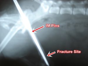
Care of Fractures
In order to repair your pet’s fracture, your veterinarian has to 1) reduce the fracture (ie. bring the ends of the break back together); 2) fix the fracture (ie. fasten fragments together); 3) immobilize the fracture (ie. prevent the fracture from moving, thereby allow the fracture to heal).
Once the fracture is repaired, you must follow your veterinarian’s advice in order to heal the fracture.
- Your pet’s exercise should be restricted. No running, jumping or rough play.
- Avoid slippery surfaces.
- Keep the cast, splint or surgery site clean and dry!
- Avoid objects or projections that may entangle the cast, splint or fixation device.
- Avoid stairs
- Monitor and keep your pet from chewing or licking the fixation device
- Contact your veterinarian immediately if a pin, wire, splint or cast is bent or broken.
- Contact your veterinarian immediately if there is a bad odor from the cast, splint or pin area.
- Contact your veterinarian immediately if the cast or splint slips down.
- Contact your veterinarian immediately if the limb is swollen or discolored.
- Contact your veterinarian immediately if your pet seems uncomfortable.
Home care is very important in successful healing of your pet’s fracture!
Cervical Spondylomyelopathy (wobbler syndrome)
Cervical spondylomyelopathy is a disorder of large and giant breeds of dogs (eg. Great Danes and Doberman Pinschers) characterized by spinal cord compression due to malformation of bony or ligamentous structures secondary to instability or chronic disc disease. Signs include pain, weakness and ataxia (difficulty walking). Diagnosis is afforded via x-rays and special dye studies (eg. myelogram). Treatment may include corticosteroids and surgery.
Cranial Cruciate Ligament Rupture
A ligament is a tough, fibrous tissue connecting bones. The cranial cruciate ligament is located in the knee and gives the knee stability when it is bent (flexion). Rupture of this ligament allows the tibia (shin bone) to slide out from under the femur (thigh bone) thereby stretching the surrounding joint capsule. Signs include pain, swelling and lameness. Left untreated the disorder will produce osteoarthritis of the knee. Diagnosis is afforded via physical exam, manipulation under anesthesia (positive cranial drawer sign) and arthroscopy. Treatment may include rest, anti-inflammatories and surgery. Surgical procedures may include imbrication, fascial strip procedure, patellar tendon procedure, tibial plateau leveling osteotomy and/or extracapsular prosthesis.
The cranial cruciate ligament is one of two ligaments within the stifle (knee joint) which pass from the femur (thigh bone) to the tibia (skin bone). These ligaments normally keep the knee “tight” and, therefore, keep the articular (gliding) surfaces of the tibia and femur in the proper position when the leg is flexed or extended. If either of these ligaments is damaged, the bones forming the knee will slide out of alignment. This instability can cause any or all of the following: inability to bear weight (lameness), pain, joint swelling, degeneration of the joint (arthritis), wobbly leg(s).
The cranial cruciate is more commonly ruptured and is more likely to cause severe gait abnormalities, immediately or within weeks to months as the arthritis worsens. Two other structures, the collateral ligaments and the menisci (cartilage), may be damaged at the same time or be damaged later due to the chronic instability of the knee joint. This happens in over 50% of the cases. (The collateral ligaments provide sideways support and the menisci serve as “shock absorbers” and gliding surfaces for the knee).
Certain body types are prone to this injury. One type is middle-aged, obese, rather inactive animals with minimal muscle development. These dogs have poor support of the knee joint and rupture is usually spontaneous (without external trauma). The other group is active, vigorous and well-muscled, and damage occurs usually in the course of athletic endeavors. In all dogs, the insult is due to some act of overextension or leg rotation.
Your dog may or may not show signs of extreme pain when this happens. Although more often unilateral, the dog may acutely collapse if both hind limbs are affected or if he cannot support full weight with only one leg. A typical history shows the dog’s weight bearing and gait abnormality gradually improving in the weeks following the initial insult. This rarely returns to normal, however, and since degenerative arthritis has begun, lameness continues.
Beside the history, the most important diagnostic test is the cranial drawer sign, whereby one can feel the tibia slide forward away from the femur. This motion is normally opposed by the cranial cruciate ligament. NOTE: If available, arthroscopy can definatively diagnose cranial cruciate ligament rupture.
Very small dogs (less than 25 pounds) may recuperate simply with strict confinement and rest for many weeks. For larger dogs, and small dogs with severe, nonresponsive signs, surgery is indicated. The goals of surgery are to: (1) regain normal stifle stability and weight bearing, and (2) remove the factors inciting arthritis.
Damaged ligaments and menisci promote arthritis. Therefore, the stifle is inspected and cleared of this debris. Methods of regaining stability are not so standardized and can include any of the following: tightening the loose tissue around the knee (imbrication), implantation of an extracapsular suture prosthesis, making a new ligament out of fibrous tissue, tibial tuberosity advancement (TTA) or tibial plateau leveling osteoplasty (TPLO).
Postoperatively, your animal will require strict exercise control for four to eight weeks. During this period, only controlled lease walking outside is allowed along with physical therapy. Uncontrolled stress on the operated leg may damage the joint.
Craniomandibular Osteopathy
Craniomandibular osteopathy is a painful disease of puppies of either sex between the ages of 4 to 10 months, predominantly in terriers (Scottish, West Highland White, Cairn) but may affect Boxers, Labrador Retrievers, Great Danes and Doberman Pinschers. The condition is neither inflammatory nor cancerous but the cause is unknown. The jaw (mandible) and adjacent bones of the skull become thickened which limits the range of jaw motion, causing pain on chewing and inability to open the mouth widely. Other signs include fever and lethargy. Diagnosis is afforded via physical exam and radiography (x-rays). Treatment is non-specific and may include analgesics and nutritional support.
Discospondylitis
Discospondylitis is an infection of one or more vertebrae and the intervertebral discs that join them. Causes include penetrating wounds, infections around the vertebrae, grass awns (eg. foxtails), blood borne bacteria or foreign materials that migrate through the body. Signs include pain, fever, reluctance to move and various neurologic signs depending on the location and severity of secondary spinal cord compression. Diagnosis is afforded via physical exam, complete blood count, blood culture, radiography and special dye studies. Treatment may include antibiotics, anti-inflammatories, surgical drainage and supportive therapy.
Elbow Dysplasia
Elbow dysplasia results from abnormal development of the ulna, one of the bones of the forearm. During growth (between 20 and 24 months of age), a small area of bone (anconeal process) fails to fuse with the rest of the bone causing an unstable elbow joint. The disorder occurs most often in young Basset hounds and German Shepherds affecting one or both front legs. The condition is hereditary, therefore affected animals should not be bred. Signs include lameness that is aggravated by exercise and secondary degenerative joint disease (arthritis). Diagnosis is afforded by physical exam and radiography. Treatment consists of surgical removal or reconstruction of the ununited fragment.
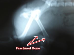
Fractures
Fractures are the breaking of bones. Fractures can be characterized in three major categories: simple fracture, where the bone breaks in two pieces; comminuted fractures, where the bone breaks into more than two pieces; compound fractures, where the broken bone breaks the skin and is therefore exposed to the outside world. Signs include pain, swelling, lameness and/or loss of normal function. Diagnosis is afforded via history, physical exam and radiography. Treatment may include rest, casting, surgical pinning and/or plating, depending on the location and extent of the lesion. For the appendages (eg. legs), the joint above and below the fracture must be immobilized. Therefore fractures above the elbow and knee must be surgically stabilized, while those below the elbow and knee might be casted.
Hip Dysplasia
Hip dysplasia is a disorder in which abnormal formation of the hip joint results in joint laxity. As the affected pet ages, the hips gradually become arthritic and may degenerate to crippling of the rear limbs. The exact cause is unknown but it is believed to develop as muscle development lags behind the rate of skeletal growth. The imbalanced growth rate is affected by heredity, diet and other unknown factors. Not all dogs are affected to the same degree as the disease can be very mild and cause no signs at all, or it may be severe and crippling. The disease is usually bilateral but occasionally affects only one side. Signs include hindleg lameness, swaying or staggering gait, “bunny-hopping”, discomfort on rising and reluctance to climb stairs, stand on the rear legs, run or jump. The disease is most common in large breeds of dogs but may occur in any breed and in cats. Diagnosis is afforded by physical exam, radiography and special manipulations under anesthesia (eg. Barlow’s, Barden’s and Ortolani signs). Treatment may include anti-inflammatories, nutrients (eg. glucosamines, chondroitin sulfates, omega 3 fatty acids), weight control, physical therapy and various surgical procedures (eg. triple pelvic osteotomy, femoral head and neck excision, pectineal myectomy, intertrochanteric osteotomy, total hip replacement) depending upon the pet’s age and severity of the disease.
Hypertrophic Osteodystrophy
Hypertrophic osteodystrophy is a bone disease of young (2-8 months old), rapidly growing large and giant breed dogs of unknown cause. The disease causes bony thickenings near the joints at the ends of the forearms and forelegs. Signs include lameness, fever, lethargy, decreased appetite and depression. Severe cases result in permanent deformed of legs. Diagnosis is afforded via physical exam and radiography. Treatment may include anti-inflammatories, analgesics, dietary restriction and good supportive care. Most dogs with hypertrophic osteodysrophy recover, but occasionally the disorder is fatal due to extremely high fever.
Hypertrophic Osteopathy
Hypertrophic osteopathy (HO) is a disease that produces a bilateral increase in the thickness of periosteum (tissue covering bone) of the limbs secondary to a space occupying mass in the abdomen or chest. Signs of HO many times occur long before those of the underlying disease and may include thickening of the distal limbs and lameness. Diagnosis is afforded via physical exam, laboratory tests and radiography. Treatment is directed at the underlying disease. Successful treatment of the underlying disease usually results in regression of the HO.
Intervertebral Disc Disease
The spine is made up of bony segments called vertebrae, which are joined by ligaments, muscles and intervertebral discs. Intervertebral discs act as shock absorbers between vertebrae and consist of a fibrous outer ring and an inner section that is soft and gelatinous. The fibrous outer ring is thinner at the top than at the bottom. When a disc becomes diseased, either through degeneration or injury, the thinner top portion of the outer ring gives way and the inner portion escapes into the spinal canal located directly above the disc. The spinal cord is located within the spinal canal and a ruptured disc causes pressure or damage to the spinal cord, resulting in pain, weakness, incoordination or paralysis. Diagnosis is afforded via physical exam, neurological tests and radiographs. The radiographs (x-rays) may include special dye injected into the spinal canal (myelography) to outline the spinal cord and any areas of damage. Treatment may include corticosteroids, surgery (decompress the area compressed with disc material) and physiotherapy.
Muscular Dystrophy
Muscular dystrophy is a sex linked, inherited disease which results in progressive muscular degeneration. The disease is caused by the expression of a mutated X-chromosome. Since males have only one X-chromosome, the recessive trait is usually expressed in males. The breeds usually affected by this rare disease include: Golden Retrievers, Cocker Spaniels, Samoyeds, Brittany Spaniels, Labrador Retrievers, Pembroke Welsh Corgies, Irish Terriers and Miniature Schauzers. Dogs with muscular dystrophy usually show signs as early as eight weeks of age. Clinical signs include muscle weakness, fatigue, exercise intolerance, stiff gait, dropping food out of their mouth and difficulty swallowing. Diagnosis is afforded by physical exam and muscle biopsy. Unfortunately there is no successful treatment. Stem cell treatment may someday successfully treat this disorder.
Osteochondrosis
Osteochondrosis is a disease of the cartilage of the shoulder, elbow, hock and/or knee joints of young, fast-growing, large-breed dogs. Lameness first appears at 6-9 months of age, predominantly in males and may wax and wane for several weeks or months. The exact cause is unknown but the condition begins as abnormal development of the deep layers of joint cartilage. As the condition progresses, a small piece of cartilage may detach from the underlying bone. Signs include pain, lameness, fever and reluctance to walk. Diagnosis is afforded via accurate history, physical exam, radiography (which may include injecting air into the joint) and arthroscopy. Treatment may include anti-inflammatories, glucosamine and chondroitin sulfate, glycosylated aminoglycans, physiotherapy and surgery (removal of cartilaginous flaps).
Osteomyelitis
Osteomyelitis is a bacterial or fungal infection of the bone usually caused by wounds, bites, open fractures, extension of existing infection and blood borne infections. Signs include swelling, fever, pain, lameness and/or drainage at the site of infection. Diagnosis is afforded via accurate history, physical exam, radiography, complete blood count, culture and sensitivity. Treatment may include antibiotics, antifungals, drainage, flushing and curettage.
Panosteitis
Panosteitis is an inflammatory bone disease primarily affecting young large breed dogs. It affects the middle portion of the long bones of one or more limbs. Contributing causes include stress, transient vascular abnormalities, allergies, metabolic disorders, elevated estrogen levels and autoimmune reactions following a viral infection. The disease is self limiting but may persist for 1-6 months, with the average case lasting 2-3 months. Signs include periods of pain and lameness, interrupted by intervals of good health, and at times switching from one leg to another. Diagnosis is afforded by accurate history, physical exam, complete blood count, blood chemistries and radiography. Treatment may include analgesics, anti-inflammatories, dietary restrictions and supportive care.
Patellar Luxation
Patellar luxation is a dislocation of the patella (kneecap). The kneecap may dislocate toward the inside (medial) or outside (lateral) of the leg and both legs may be affected. Causes include trauma or congenital (present at birth) deformities. The effects of patellar luxation are related to the severity and duration of the luxation. Signs include pain and lameness which may be intermittent but milder forms, especially in small breeds, may show little or no signs. Diagnosis is afforded via physical exam (at times aided by sedation). Treatment may include nothing (with mild case without clinical signs of lameness) or surgery (patellar arthroplasty).
Rheumatoid Arthritis
Rheumatoid arthritis is an uncommon immune-mediated joint disease that usually affects small or toy breeds of dogs. Onset of the disease is usually around 4 years of age although any age can be affected. The cause is probably due to a defect in the pet’s immune system. Signs may include a shifting leg lameness, joint swelling, episodic fever, lethargy and limb deformity with prolonged disease. Diagnosis is afforded via physical exam, complete blood count, radiographs, joint fluid analysis and laboratory tests (eg. antinuclear antibody, rheumatoid factor). Treatment may include glucocorticoids, anti-inflammatories, immune suppressive drugs and supportive care.
Rickets
Rickets is a disease of young, growing animals characterized by improper bone development. The disease is caused by a dietary imbalance of calcium, phosphorus and vitamin D. Signs include swelling of the joints, bending or bowing of the long leg bones and fractures. Diagnosis is afforded by accurate history, physical exam, blood chemistries and radiographs. Treatment includes dietary correction and supportive care. Because most dogs are fed commercial dog foods that are properly balanced, the disease is not common yet improper mineral supplementation or a diet high in meats can manifest rickets.
Spinal Disorders
The spinal column (vertebrae) extends from the base of the skull through the tail. It gives support to the body and protects the spinal cord from which nerve branches stem out to reach vital structures and muscles of the body. Fractures, arthritis and various other diseases of the spinal column may cause disorders, ranging from pain to paralysis. Neurologic exams and radiographs (x-rays) may be necessary to evaluate your pet’s condition.
Sprains
Sprains are injuries to joints, tendons or ligaments. Though sprains may be very painful, bones are not broken and the lameness is usually temporary. Injuries of this sort are common and occur more in young or very energetic pets.
Umbilical Hernia
An umbilical hernia is the protrusion of abdominal contents through the body wall, beneath the skin at the naval (umbilicus). The umbilicus is the”belly button” in the mid abdominal area marking the opening through which the prenatal blood vessels and other fetal structures passed before birth. After the umbilical cord is broken/cut at birth, the opening rapidly closes. Occasionally it does not close completely and an opening in the abdominal wall remains. This presents the danger of intestines passing through the opening and becoming entrapped. If this occurs, the intestinal blood supply may be strangled, the passage of bowel content may be blocked and intestinal tissue may die. This releases deadly toxins producing shock and possible death of the pet.


